The Renal Pelvis: Your Kidney's Urine Highway - Explained
Why is the renal pelvis so crucial to the function of your kidneys? The renal pelvis serves as a critical conduit, directing the flow of urine from the kidney to the bladder, and its health is essential for overall urinary system function.
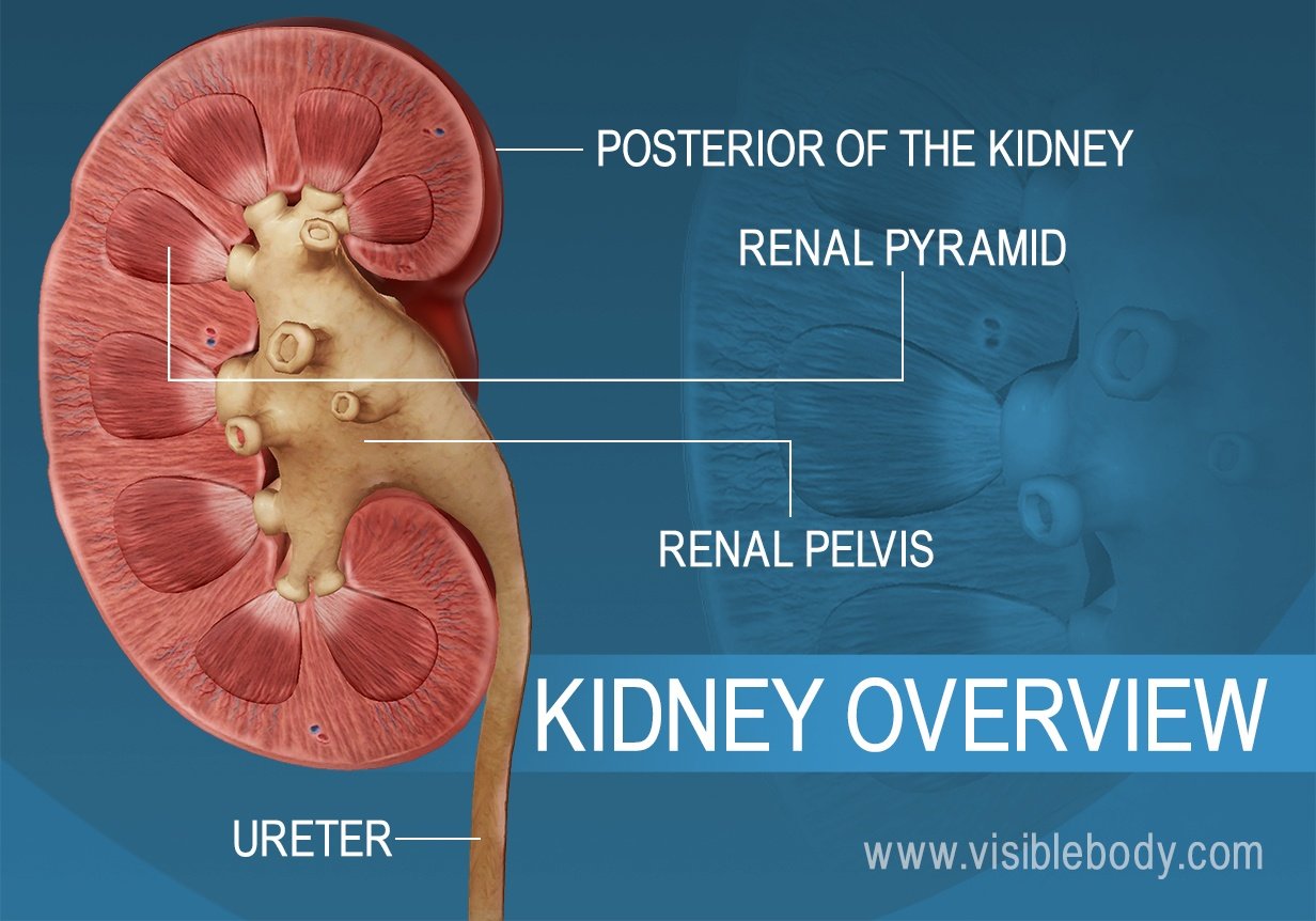
The renal pelvis, an often-overlooked yet vital component of the urinary system, plays a pivotal role in the complex process of waste filtration and removal. Its structure and function are intricately linked to the overall health of the kidneys and the efficient elimination of urine from the body. Located within the medial, concave surface of the kidney, it's an expanded, funnel-shaped area that serves as the primary collection point for urine produced within the kidney itself. This crucial anatomical feature is not merely a passive conduit; it actively participates in the transportation of urine, ensuring its smooth passage to the ureter and, ultimately, the bladder.
The renal pelvis isn't an isolated entity; it's an integral part of a larger system designed for waste removal. Within the kidney, the formation of urine begins in the nephrons, the microscopic functional units responsible for filtering blood. As urine is produced by these nephrons, it flows into the renal pyramids, structures located within the renal medulla. From the renal pyramids, the urine is channeled into the renal papillae, the apex of the renal pyramids, which then drain into the minor calyces. These minor calyces merge to form the major calyces, and it is at this juncture that the renal pelvis comes into play. The major calyces empty into the renal pelvis, a central, funnel-shaped space that funnels the urine into the ureter.
The architecture of the renal pelvis is designed for efficiency and resilience. Its inner surface is lined with a specialized tissue known as transitional epithelium, a stretchy, multi-layered tissue that allows the renal pelvis to expand and contract as urine flows through. The wall of the renal pelvis also comprises smooth muscle fibers, arranged in a longitudinal fashion. These muscle fibers facilitate peristalsis, a wave-like contraction that propels urine towards the ureter, ensuring its continuous flow. The apex of the renal pelvis extends outwards from the kidney and seamlessly merges with the superior end of the ureter, the muscular tube that carries urine to the bladder. The renal pelvis contains the hilum.
Understanding the anatomy of the renal pelvis is essential for grasping how the kidneys function. A kidney diagram can be a powerful tool for visualizing the intricate network of structures within the kidney. By identifying key parts such as the renal cortex, medulla, and pelvis, one can gain valuable insight into the processes that filter waste, regulate fluid balance, and maintain overall health. The renal cortex, the outer layer of the kidney, contains the nephrons, the functional units of the kidney, which filter the blood. The renal medulla, the inner layer, is home to the renal pyramids, cone-shaped structures that collect urine from the nephrons. The renal pelvis then collects this urine and channels it into the ureter.
The renal pelvis is not only crucial for the normal functioning of the urinary system but also plays a role in various medical conditions. It is the location of several types of kidney cancer, and it is affected by infections such as pyelonephritis, a bacterial infection of the kidney. Furthermore, the size of the renal pelvis is a critical factor in grading hydronephrosis, a condition characterized by the swelling of the kidney due to a backup of urine. Staghorn kidney stones, large stones that can fill the renal pelvis and its branches, may block all or part of the renal pelvis, leading to significant complications. Any disruption to the renal pelvis's function can have serious consequences for the urinary system.
The journey of urine through the renal pelvis is a meticulously orchestrated process. Urine, formed by the nephrons within the renal pyramids, is funneled into the minor calyces, which then converge to form the major calyces. These major calyces then empty into the renal pelvis. The smooth muscle within the renal pelvis contracts in a coordinated fashion, propelling the urine into the ureter. This rhythmic contraction, known as peristalsis, ensures a steady flow of urine towards the bladder. From the renal pelvis, urine flows into the ureter, a muscular tube that transports the urine to the urinary bladder for storage until voiding. The connection between the renal pelvis and the ureter is seamless, ensuring the uninterrupted flow of urine. The renal pelvis receives about 2 major calyces, which in turn receive minor calyx, which in turn collect urine from the papilla. It functions as a pathway for fluid on its way to the bladder.
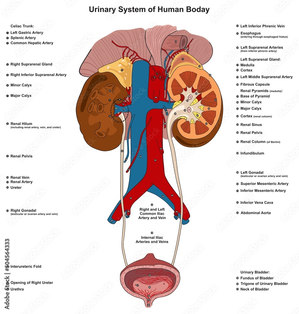
The renal arteries, which supply blood to the kidneys, form directly from the descending aorta. In contrast, the renal veins, which carry cleansed blood away from the kidneys, drain directly into the inferior vena cava. This strategic placement of blood vessels ensures efficient filtration and waste removal. The blood vessels that supply the kidney also play a role in the anatomy, the most important blood vessels entering and exiting the kidneys are the renal artery and renal vein. The diagram of the urinary system shows the organs responsible for producing, storing, and excreting urine from the body. The diagram of the urinary system of humans shows that it is made of kidneys, ureters, bladder, and urethra. It also includes additional structures like renal arteries and veins, which supply blood to the kidneys and adrenal glands.
The renal pyramids are separated by extensions of the cortex called the renal columns. Each renal papilla is associated with a structure known as the minor calyx, which collects urine from the pyramids. The minor calyces converge to form two or three larger drains called major calyces, which eventually converge to a structure called the renal pelvis. The renal pelvis is connected to the ureter. The smooth muscle in the renal pelvis funnels urine via peristalsis into the ureter. From the renal pelvis, the urine passes into the ureter.
The renal medulla contains the renal pyramids, where urine formation takes place. Each pyramid creates urine and terminates into a renal papilla. The pyramids are separated by extensions of the cortex called the renal columns. The central renal sinus, consisting of the calyces, renal pelvis and fat, is more echogenic than the cortex. The renal pelvis may appear as a central slit of anechoic fluid at the hilum. The renal pelvis leads to the ureter on the outside of the kidney.
Internally, the kidneys consist of 2 layers; a highly vascularized outer renal cortex and an inner renal medulla. Spanning across these two layers are millions of the kidneys. The cavity of the pelvis and calyx surrounds the renal papilla (or its equivalent in other kidney types). The minor calyces converge to form two or three larger drains called major calyces, which eventually converge to a structure called the renal pelvis. Learn more about the renal pelvis in this article. The renal pelvis is connected to the ureter. It functions as a pathway for fluid on its way to the bladder.
The adrenal glands sit on the superior pole of each kidney. Urine is collected into a system of renal calices, which is a series of distinctive chambers within a kidney. Calices gradually increase in size, starting with the minor calices, which open into larger major calices, which empty into the renal pelvis. The renal pelvis collects the urine and channels it into the ureter.
The diagram of urinary system shows the organs responsible for producing, storing, and excreting urine from the body. It also includes additional structures like renal arteries and veins, which supply blood to the kidneys and adrenal glands. Each kidney contains around one million nephrons, which are the functional units of the kidney. Usually, there are two to three major calices in the kidney (superior, middle, and inferior), which again unite to form the renal pelvis from which the ureter emerges and leaves the kidney through the hilum. The apex of a renal pyramid is called a renal papilla. Several minor calices merge to form a major calyx.
The renal sinus, consisting of the calyces, renal pelvis and fat, is more echogenic than the cortex. Normal ureters are generally not well seen on ultrasound. On unenhanced ct the renal pyramids can appear hyperdense. The renal pelvis of the dog kidney. Finally, the collected urine passes into the ureter through the renal pelvis of the dog. The renal pelvis of a dog is elongated in a craniocaudal direction. The renal pelvis connects the kidney to the rest of the body. Urine is collected into a system of renal calices, which is a series of distinctive chambers within a kidney. Calices gradually increase in size, starting with the minor calices, which open into larger major calices, which empty into the renal pelvis. From the renal pelvis, the urine passes into the ureter. They channel urine from the pyramids to the renal. The minor calyces converge to form two or three larger drains called major calyces, which eventually converge to a structure called the renal pelvis.
The smooth muscle in the renal pelvis funnels urine via peristalsis into the ureter. The renal arteries form directly from the descending aorta, whereas the renal veins return cleansed blood directly to the inferior vena cava. The renal pelvis is divided into calyces. Each pelvis receives about 2 major calyces, which in turn receive minor calyx, which in turn collect urine from the papilla. The adrenal glands sit on the superior pole of each kidney. The diagram of urinary system shows the organs responsible for producing, storing, and excreting urine from the body. The diagram of the urinary system of humans shows that it is made of kidneys, ureters, bladder, and urethra. It also includes additional structures like renal arteries and veins, which supply blood to the kidneys and adrenal glands. A frontal section through the kidney reveals an outer region called the renal cortex and an inner region called the renal medulla. Each pyramid creates urine and terminates into a renal papilla. The most important blood vessels entering and exiting the kidneys are the renal artery and renal vein.
From the renal pelvis, the urine passes into the ureter. 1a and 1b) or the renal calyces are anchored to the renal parenchyma by connective and smooth muscle tissues that follow the intrarenal arteries. The cavity of the pelvis and calyx surrounds the renal papilla (or its equivalent in other kidney types). Each kidney contains around one million nephrons, which are the functional units of the kidney. The renal pelvis is divided into calyces. Each pelvis receives about 2 major calyces, which in turn receive minor calyx, which in turn collect urine from the papilla. It functions as a pathway for fluid on its way to the bladder.
The renal pelvis is an expanded funnel shaped area through which urine travels. It is located within the medial, concave surface of the kidney, filling the renal sinus. The apex of the renal pelvis extends outwards from the kidney, and becomes continuous with the superior end of the ureter. Urine is formed inside the kidney by nephrons. By identifying key parts of the kidneys, such as the renal cortex, medulla, and pelvis, you can gain valuable insight into how these organs work to filter waste, regulate fluid balance, and maintain overall health. The renal pelvis is the location of several kinds of kidney cancer and is affected by infection in pyelonephritis. a large staghorn kidney stone may block all or part of the renal pelvis. The size of the renal pelvis plays a major role in the grading of hydronephrosis. Renal pelvis, enlarged upper end of the ureter, the tube through which urine flows from the kidney to the urinary bladder. The pelvis is almost completely enclosed in the deep indentation on the concave side of the kidney, the sinus. It forms at the junction of the major calyces. It is the region where urine from different parts of the kidney comes together. The inner surface of the renal pelvis is lined with a stretchy tissue called transitional. The wall of the renal pelvis consists of longitudinal smooth muscle fibers. This enables a milking function of urine from the renal pelvis into the ureter. On radiographs, the ureter is divided into three sections. Emerging from the hilum is the renal pelvis, which is formed from the major and minor calyxes in the kidney. The smooth muscle in the renal pelvis funnels urine via peristalsis into the ureter. The renal arteries form directly from the descending aorta, whereas the renal veins return cleansed blood directly to the inferior vena cava. There are, on average, eight renal pyramids in each kidney. The renal pyramids along with the adjoining cortical region are called the lobes of the kidney. The renal pelvis leads to the ureter on the outside of the kidney. On the inside of the kidney, the renal pelvis branches out into two or three extensions called the major calyces, which. The renal medulla contains the renal pyramids, where urine formation takes place. The pyramids are separated by extensions of the cortex called the renal columns. Each renal papilla is associated with a structure known as the minor calyx, which collects urine from the pyramids. Several minor calices merge to form a major calyx.
| Renal Pelvis: Essential Information | |
|---|---|
| Definition: | The expanded, funnel-shaped upper end of the ureter within the kidney. |
| Location: | Medial, concave surface of the kidney; within the renal sinus. |
| Function: | Collects urine from the major calyces and funnels it into the ureter for transport to the bladder. |
| Structure: | Lined with transitional epithelium and contains smooth muscle fibers for peristalsis. |
| Clinical Significance: | Affected by kidney stones, infections (pyelonephritis), and kidney cancers; plays a role in hydronephrosis grading. |
| Connected Structures: | Receives urine from major calyces and connects to the ureter. |
| Additional Points: |
|
| Reference: National Center for Biotechnology Information (NCBI) |
The kidneys, complex organs responsible for filtering waste and maintaining fluid balance, rely heavily on the renal pelvis for the efficient removal of urine. This vital structure, along with the other components of the urinary system, works in harmony to ensure the proper functioning of the body. The renal pelvis serves as a critical bridge, facilitating the smooth transition of urine from the kidney to the bladder, thus ensuring that waste products are effectively eliminated. Anomalous position of the renal hilum. The hilum is the point of entry and exit for blood vessels, nerves, and the ureter.
Urine passes from the renal pyramids into the renal pelvis. Urine drains from the renal pelvis into the ureter. (a)renal sinus (b)renal pelvis (c)hilum (d)renal papilla (e)ureter (f)renal cortex (g)renal medulla (h)renal pyramid (i)minor calyx (j)major calyx (k)kidney lobe (l)renal columns (m)fibrous capsule the basic functional unit of the kidney is the nephron. A frontal section through the kidney reveals an outer region called the renal cortex and an inner region called the renal medulla. Each pyramid creates urine and terminates into a renal papilla. The most important blood vessels entering and exiting the kidneys are the renal artery and renal vein. The adrenal glands sit on the superior pole of each kidney. It is thin but tough and fibrous. They channel urine from the pyramids to the renal. The minor calyces converge to form two or three larger drains called major calyces, which eventually converge to a structure called the renal pelvis. The renal pelvis is connected to the ureter. The renal pelvis collects the urine and channels it into the ureter.

